Accurate understanding of dental morphology is fundamental to both veterinary anatomical education
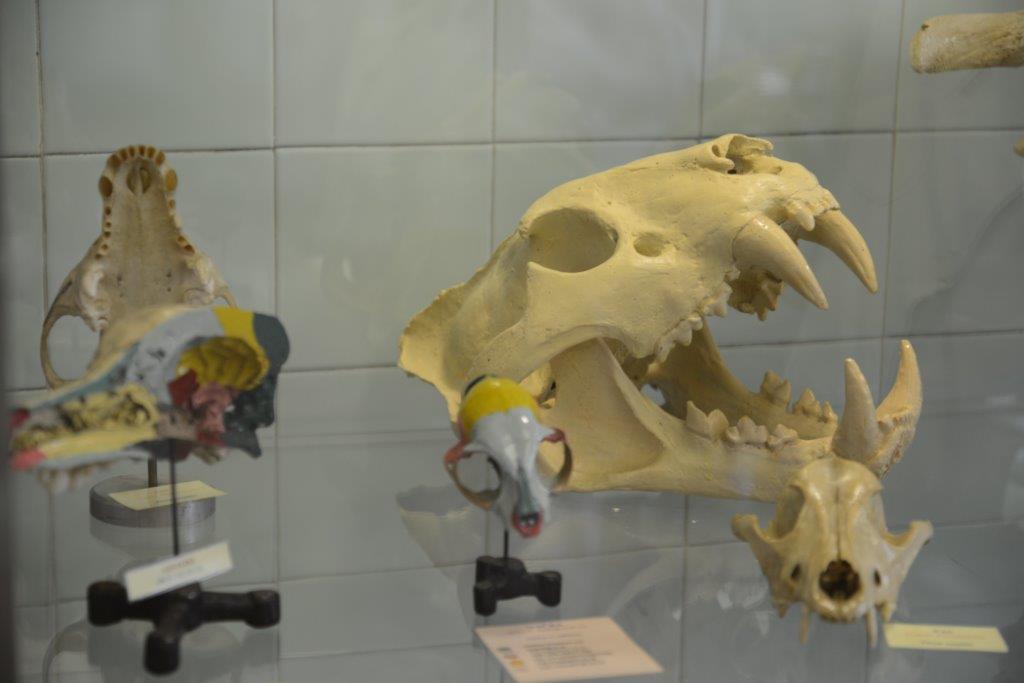
and clinical decision-making. This hands-on workshop provides participants with the opportunity to explore and compare the dentition of a wide range of domestic and wild animal species using real skulls and extracted teeth. Attendees will handle individual teeth and practice placing them into their corresponding alveoli, reinforcing their ability to identify species-specific dental characteristics, crown shape, root configuration, and functional adaptations.
To highlight the clinical relevance of dental anatomy, the workshop also includes skulls with marked cutting lines illustrating recommended approaches for tooth extraction. This allows participants to visualise anatomical landmarks, anticipate challenges posed by variable root morphology, and appreciate how detailed anatomic knowledge directly informs safe and effective dental procedures.
By combining comparative anatomy with practical clinical demonstrations, this workshop aims to deepen participants’ understanding of dental structures, enhance their diagnostic confidence, and strengthen their surgical planning skills in veterinary dentistry.